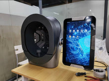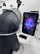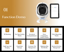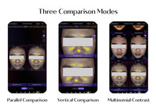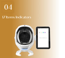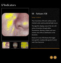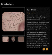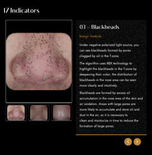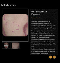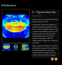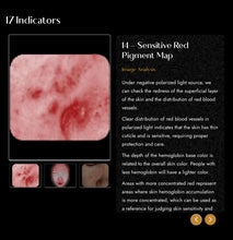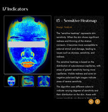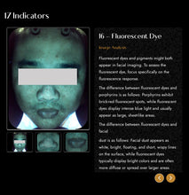
 Follow the road map to comprehensive and accurate 21st Century skincare with the state of the art JuvaMap AI 17 Indicator 3D Skincare Analyzer!
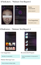
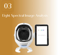
Designed for precision skin analysis, this innovative device integrates eight spectral imaging technologies to detect 17 facial skin concerns with ease.
The cutting edge JuvaMap Skincare Analyzer easily and quickly provides insight into your skin to detect common and unnoticeable skin problems such as skin concerns, including dark circles, radiance, redness, pores, wrinkles, acne, and more.
This essential skincare tool identifies all skin types including tan skin and performs a skin comprehensive age test, and provides personalized skincare routine recommendations with 21st Century AI Software so your skin looks flawless and you won’t waste money on unnecessary products.
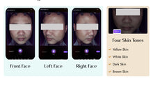
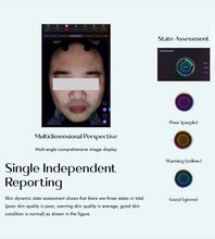
With the patented ‘Smart Scan’ 3D AI Software, the JuvaMap Skin Analyzer quantitatively , qualitatively, and precisely analyzes multiple skin problems in real time using state of the art multi spectrum LED imaging technology.
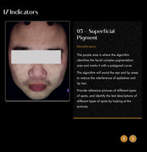
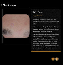
This includes pigments, deep spots, sensitivity, wrinkles, pores, acne, black and whiteheads, and superficial vascular and fluorescent concerns.
Perfect for homes, dorm rooms, professional offices, the easy to use touchscreen technology captures consistent views with ease using our multi-point positioning system and live image overlay for accurate analysis and documentation of your progress over time.
How it Works:
Using a proven combination of advanced Artificial Intelligence (AI) to detect Acne activity level together with medical expert approved skin indicators to help identify your skin type and acne type.
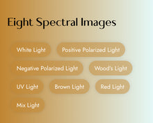
 The advanced Skin Analysis System uses eight distinct light sources, including natural sunlight, cross-polarized light, UV light, and parallel polarized light, plus an impressive 35 million pixels to accurately and comprehensively reveal skin conditions.
- Daylight mode: This mode enables users to examine a customer's skin in a controlled environment with natural light. This allows for comparison of skin conditions in daylight mode with other analysis modes. In particular, the system effectively analyzes pores, wrinkles, and skin color in daylight and other lighting conditions.
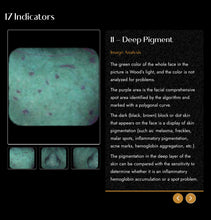
- Cross-polarized light mode: This mode reduces surface glare of the skin, allowing for detailed inspection of skin issues such as sensitivity or abnormal pigmentation.
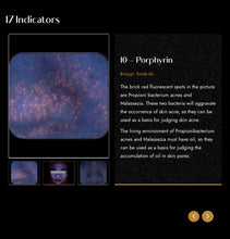
- UV light mode: By analyzing the various fluorescence performances and secretions of skin cells, the UV light mode allows you to accurately identify challenging skin conditions that may not be easily noticeable or distinguishable. This includes issues such as pigmentation and porphyrin levels.
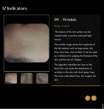
- Parallel polarized light source: In parallel polarized light mode, specular reflection is improved, making it easier to identify skin surface issues. Its main purpose is to examine fine lines, pores, and other skin surface imperfections.
- White Light: When applied to skin analysis, RGB white light can provide valuable information about skin tone, texture, pigmentation, and overall appearance
- Blue Light: When applied to skin analysis, Blue light can provide valuable information about various aspects of the skin’s health and appearance.
- Woods Light: When applied to skin analysis, Wood’s light can provide valuable information about pigmentation, acne, and sebum levels.
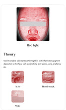
- Red Light:Red light imaging, also known as reflectance imaging utilizes red light to illuminate the skin’s surface and capture reflected light, providing valuable information about the skin’s structure, texture, pigmentation, and more.
Want to know the age of your skin?
With this cutting-edge technology you can zoom in on your pores to see their depth and size.
The instrument is very helpful in managing and tracking your progress to see what is working well and avoiding mistakes to help you accurately and quickly achieve your goals!
The technology can detect both superficial skin problems like wrinkles and the ones hidden skin base layer like oil secretion through the quantitative analysis.
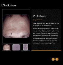
The instrument allows you to completely understand your facial skin spots, flare, chloasma, red blood silk, wrinkles, loss of collagen, skin texture, large pores, acne .
The test reveals your true skin type and identifies underlying causes of skin conditions you might have such as breakout activity, dehydration, sensitivity or uneven skin tone.
FAQ’s
What are the definitions of the various skin features and how are the features detected?
Spots : Spots are typically brown or red skin lesions including freckles, acne scars, hyper-pigmentation and vascular lesions. Spots are distinguishable by their distinct color and contrast from the background skin tone. Spots vary in size and generally have a circular shape.
Pores : Pores are the circular surface openings of sweat gland ducts. Due to shadowing, pores appear darker than the surrounding skin tone and are identified by their darker color and circular shape. The system distinguishes pores from spots based on size; by definition, the area of a pore is much smaller than a spot.
Wrinkles : Wrinkles are furrows, folds or creases in the skin, which increase in occurrence as a result of sun exposure, and are associated with decreasing skin elasticity. This skin feature has the greatest variability from image to image as it is highly dependent upon the facial expression of the client. Wrinkles are identified by their characteristic long, narrow shape.
Texture :Texture is primarily an analysis of skin smoothness. Texture measures skin color and smoothness by identifying gradations in color from the surrounding skin tone, as well as peaks (shown in yellow) and valleys (shown in blue) on the skin surface that indicate variations in the surface texture.
Porphyrins : Porphyrins are bacterial excretions that can become lodged in pores and lead to acne. Porphyrins fluoresce in UV light and exhibit circular white spot characteristics.
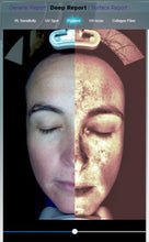
UV Spots : UV spots occur when melanin coagulates below the skin surface as a result of sun damage. UV spots are generally invisible under normal lighting conditions. The selective absorption of the UV light by the epidermal melanin enhances its display and detection by the instrument .
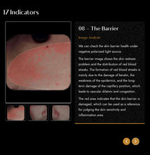
Red Areas : Red Areas represent a potential variety of conditions, such as acne, inflammation, Rosacea or spider veins. Blood vessels and hemoglobin contained in the papillary dermis, a sub-layer of skin, give these structures their red color, which is detected by the Technology. Acne spots and inflammation vary in size but are generally round in shape. Rosacea is usually larger and diffuse compared to acne, and spider veins usually are short, thin and can be interconnected in a dense network.
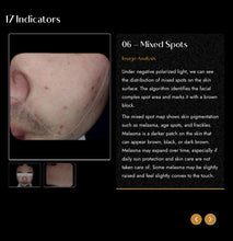
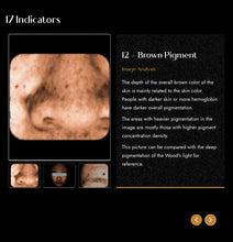
Brown spots: Brown Spots are lesions on the skin such as hyper-pigmentation, freckles, lentigines, and melasma. Brown Spots occur from an excess of Melanin. Melanin is produced by melanocytes in the bottom layer of the epidermis. Brown Spots produce an uneven appearance to the skin, and are detected.
 Features:
✔4K camera with 35 milion high-definition pixels. Demonstrates clear images that can be adjusted and magnified
✔Comprehensive skin analysis across 12 spectrums addresses various skin issues
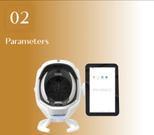
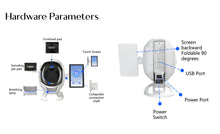
✔13.3-inch integrated touchscreen android tablet
✔True 3D thermal imaging displays high-resolution heat patterns locally.
✔Al Analysis of Over 30 skin Indicators
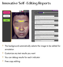
✔️ Get a detailed personal skin report in under 3 minutes
✔️Understand your skin and get personal solutions
Specs:

What’s in the box:
4K Camera 1X
13.3” Android Tablet 1X
Smart Scan Software 1X
Power Supply 1X
Warranty: 1 Year


